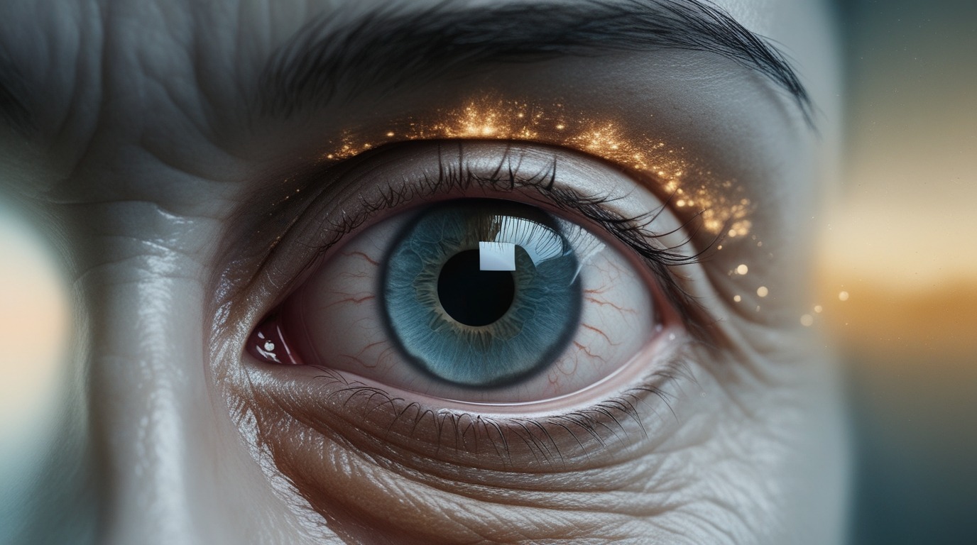
A disorder called glaucoma damages the visual nerve and can result in irreversible blindness. While it is a severe eye disease, its development can be controlled effectively through the use of some treatment options like medicated eye drops as well as surgery.
Glaucoma is quite common, especially those who are already aged 60 years old and above. In fact, white people are more likely to get Glaucoma after 60 and the risk for black or Hispanic might begin in their 40s.
The most common form of this disease, Open-angle glaucoma, comes on slowly and usually doesn’t come with symptoms at first. The thing is, early detection is possible in these cases with the help of regular eye exams.
While there is currently no known cure for glaucoma, early discovery can stop the condition from worsening or at least slow its course.
This article covers the causes, symptoms and treatment approaches of glaucoma. We’ll also discuss the different types of glaucoma and possible surgical interventions as well.
What is Glaucoma?
Glaucoma refers to a condition that affects the optic nerve, often resulting from increased pressure within the eye.The optic nerve, which connects the eye to the brain, allows us to interpret what we see.When this nerve is damaged, it affects vision, typically starting with peripheral vision and, if left untreated, can lead to total blindness.
A build-up of pressure inside the eye that damages the optic nerve is referred to as glaucoma.This damage is often caused by abnormally high pressure in the eye, also known as intraocular pressure (IOP).
The front of the eye has a clear fluid inside it called aqueous humor. It also fills the eye and helps maintain its form. Evolutionarily, the eye continually produces this liquid to reduce dehydration but it drains from a drainage system.
In glaucoma, the fluid (known as aqueous humor) drains too slowly from the eye causing pressure to build up. During a few types of glaucoma, the drainage tract ( which is the space between the iris (the colored part of the eye) and the cornea (the transparent outer layer) becomes blocked. This suggests liquid cannot flow out properly in your eye. fluid builds occurring on undesirable pressure within the eye raises at this reduction.
If a person can’t handle this pressure it could destroy the optic nerve and other parts of the eye causing vision loss also.
Both eyes are often affected by glaucoma, though one may be more severely affected.
Causes and risk factors
Although the precise origin of glaucoma is unknown, elevated intraocular pressure (IOP) is typically linked to the condition. When aqueous humor (eye fluid) is overproduced or improperly drains, pressure builds up inside the eye, leading to glaucoma.
There is no known cause of primary glaucoma in humans.If they have secondary glaucoma, there’s an underlying cause,which could be a tumor, diabetes,hypothyroidism,an advanced cataract or inflammation.
Some risk factors for glaucoma include:
Genetics: The risk is increased by a family history of glaucoma.
Age: Persons over 60 are more vulnerable.
Ethnicity: People of Asian, Hispanic, and African American descent are particularly vulnerable.
Medical conditions: Diabetes, hypertension, and nearsightedness can increase risk.
Prolonged corticosteroid use: Extended use of corticosteroid medications can elevate IOP.
- for Caucasian individuals, being older than 60
- for Hispanic and Black individuals, being older than 40
- having diabetes or a different underlying medical disease
- an ancestry of glaucoma
- having an eye condition or injury
- prior ocular surgery
- extreme nearsightedness, or myopia
- using corticosteroid drugs, particularly eye drops
- elevated blood pressure
- hereditary factors that can cause glaucoma in children
Types of Glaucoma:
Glaucoma is classified into several types depending on the mechanism of increased intraocular pressure and how it affects the eye.
- Primary Open-Angle Glaucoma (POAG)
- Angle-Closure Glaucoma
- Normal-Tension Glaucoma ( low-tension glaucoma)
- Childhood Glaucoma
- Secondary Glaucoma
Primary Open-Angle Glaucoma (POAG)
This type of glaucoma is the most common. It happens when the eye’s drainage ducts gradually clog, raising intraocular pressure (IOP) over time. Damage to the optic nerve is the outcome of the disorder, which advances without pain or early symptoms.
Angle-Closure Glaucoma
This kind develops when the drainage angle that normally connects the iris and cornea gets clogged, leading to an abrupt rise in intraocular pressure.It can be a medical emergency that requires immediate attention.
- Acute Angle-Closure Glaucoma: Characterized by a sudden and severe increase in eye pressure, with symptoms such as intense eye pain, nausea, and blurred vision.
- Chronic Angle-Closure Glaucoma: Develops gradually with a slow increase in pressure, often with less dramatic symptoms compared to acute angle-closure glaucoma.
Secondary Glaucoma
Other medical disorders like inflammation, eye injuries, or the use of certain drugs that raise eye pressure can cause this kind of glaucoma.
- Pigmentary Glaucoma: brought on by iris pigment particles obstructing the drainage canals.
- Exfoliative Glaucoma: Results from flaky material from the lens blocking the drainage system.
- Neovascular Glaucoma: Triggered by abnormal blood vessel growth over the drainage area, often associated with conditions like diabetes.
Normal-Tension Glaucoma
Even when intraocular pressure (IOP) is within the usual range, optic nerve damage can occur in normal-tension glaucoma, sometimes referred to as low-tension glaucoma. Intraocular pressure typically falls within a standard range of 10-21 mmHg, but in normal-tension glaucoma, optic nerve damage and visual field loss occur even when eye pressure is considered normal.
- Low-Tension Glaucoma: A subtype of normal-tension glaucoma characterized by the same features of optic nerve damage occurring despite normal eye pressure. The management approach is similar to that of normal-tension glaucoma.
Childhood Glaucoma (also known as Pediatric Glaucoma)
IT is a rare condition that Occurs in babies and young children, due to developmental abnormalities in the eye’s drainage system. It can be congenital or develop in the early years of life, and may lead to vision problems if not treated promptly.
The young person could have:
- abnormally big eyes
- excessive tearing
- haziness within the cornea
- light sensitivity
- Congenital Glaucoma: Present at birth, caused by developmental issues in the eye’s drainage system.
- Infantile Glaucoma: takes place in the first several years of life.
- Juvenile Glaucoma: usually after the age of three in older youngsters.
Each type and subtype of glaucoma requires specific management and treatment strategies to prevent vision loss and preserve eye health.Effective therapy depends on early detection and routine eye exams.
Symptoms of Glaucoma
Glaucoma is known as the “silent thief of sight” because it often shows no symptoms in the early stages. The following warning signs and symptoms may manifest as the condition worsens:
Open-angle glaucoma symptoms:
It’s possible that symptoms won’t become apparent until later on because they grow gradually.
Among them are:
- Gradual loss of peripheral vision
- Tunnel vision in advanced stages
Angle-closure glaucoma symptoms:
Symptoms of acute glaucoma include those that appear suddenly and include:
- Severe headache
- Eye pain
- Blurred vision
- Halos around lights
- Nausea and vomiting
- Redness in the eye
Diagnosis of Glaucoma
Regular eye exams are crucial for early detection of glaucoma, especially for those at high risk. The following diagnostic tests are commonly used:
Tonometry (Measuring IOP)
Following the application of eye drops to numb the eye, the physician employs an apparatus that either contacts the cornea (applanation) or inflates the eye to measure the pressure inside the eye.
Ophthalmoscopy (Optic Nerve Examination)
The ophthalmologist puts drops to the eye to enlarge the pupil, and then uses a special light and magnifying glass to inspect the inside of the eye.
Perimetry (Visual Field Test)
To examine the patient’s peripheral (side) vision, the physician performs a visual field exam. While the doctor delivers a bright spot at various points along the periphery of the patient’s eyesight, the patient looks straight ahead. In doing so, a map of what the person can see is created.
Gonioscopy (Examining the Drainage Angle)
The doctor numbs the damaged eye with eye drops and then covers it with a type of contact lens. A mirror embedded in the lens can display if the angle formed by the iris and cornea is normal, excessively broad (open), or excessively narrow (closed).
Pachymetry (Measuring Corneal Thickness)
The doctor inserts a probe into the front portion of the eye to measure the thickness of the cornea. Since corneal thickness might alter readings of ocular pressure, the physician will consider this when evaluating all the data.
Each of these tests is required in order to make the diagnosis of glaucoma. When combined, they offer a thorough perspective.
Treatment for Glaucoma
The goal of treatment is to either decrease the amount of fluid produced in the eye or increase its outflow and stop more damage to the visual nerve.
There are various methods for doing this:
Medications
Eye drops: Eye drops are typically used as a first line of treatment. These either increase drainage or decrease the amount of fluid produced by the eye.
For the best outcomes and to avoid negative consequences, it is crucial to carefully follow a healthcare provider’s directions.
Eye drops include, for example:
- prostaglandins
- ATP-binding enzyme inhibitors
- cholinergic substances
- beta-blockers
- releasers of nitric oxide
- rho kinase blockers
Among the negative impacts are:
- Redness
- stinging
- hue shift of the eyes color or the skin surrounding
- headaches
- sporadically, breathing difficulties or retinal detachments
- dry mouth
The doctor can advise switching to a different drug or altering the dosage if the side effects don’t go away.
Oral medications: If eye drops are insufficient to lower ocular pressure, doctors may occasionally prescribe oral medicines.
Laser Therapy
Laser treatments can help increase fluid outflow or reduce fluid production:
Trabeculoplasty: A laser is used to enhance the drainage system in cases of open-angle glaucoma.
Iridotomy: To enable fluid drainage in cases of angle-closure glaucoma, a tiny hole is created in the iris.
Surgery for Glaucoma
A surgeon could advise surgery if medication and laser therapy is not helpful or the patient is unable to take them.
Reducing intraocular pressure is often the goal of surgery.
Among the potential remedies are:
Trabeculectomy: In order to facilitate the flow of fluid out of congested drainage tubes, the surgeon uses a laser beam.
Glaucoma Drainage Implants: If glaucoma develops in childhood or as a result of another medical problem, this might be helpful. To enhance drainage, the surgeon places a tiny silicone tube into the eye.In short A small tube is inserted to help drain excess fluid.
Minimally Invasive Glaucoma Surgery (MIGS): Compared to traditional surgeries, these more recent, less intrusive methods have fewer dangers and can help relieve ocular pressure.
Filtering surgery: In order to enhance fluid outflow, the surgeon may create channels in the eye if laser surgery is ineffective.
Treating acute angle-closure glaucoma
An acute angle closure in glaucoma patients is a medical emergency.
Medication to reduce hypertension will be administered right away by a doctor.
They might employ a laser treatment to make a tiny hole in the iris that would let fluids enter the drainage system of the eye.This process is known as an iridotomy.
Even if there is a potential that the second eye may also acquire glaucoma, the doctor may treat both eyes if one has glaucoma.
Prevention
Even though there isn’t a known cure for glaucoma, early identification and treatment can typically prevent visual loss.
Regular ocular examinations are crucial, as they are the only means of identifying glaucoma in its early stages.The Glaucoma Foundation recommends a baseline test at forty years of age. The results will help the doctor identify any changes in the future.
A physician can provide guidance on the frequency of eye exams based on a patient’s risk assessment.
Summary
In summary, as people age, they are more likely to get glaucoma, a frequent eye condition. It occurs when fluid in the eye does not drain, which raises the pressure and puts the optic nerve at danger of injury.
In its early stages, it might not show any symptoms, but it can cause visual loss. In order to initiate treatment, usually with eye drops, a person can start with regular eye examinations to help detect changes. This treatment can halt or delay vision loss.


1 thought on “Know Everything About Glaucoma Symptoms, Risk Factors and Treatment”
Comments are closed.