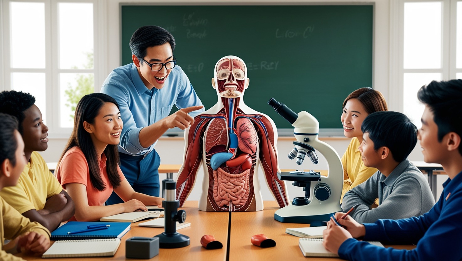
The identification and description of the structures of living things is called deconstruction, or anatomy.It’s a branch of biology and medicine. People who study deconstruction (Anatomy) study the body, how it’s made up, and how it works.
The study of deconstruction (Anatomy) dates back further than 2,000 years, to the Ancient Greeks. There are three broad areas:
- human mortal deconstruction (Anatomy)
- animal deconstruction — zootomy
- plant deconstruction — phytotomy
Anatomy, or the dissection of the human body, is the study of the body’s structures. An understanding of deconstruction (Anatomy) is crucial to the practice of drugs and other areas of health.
“Deconstruction (Anatomy)” is derived from the Greek terms “book,” which means “a slice,” and “corpus,” which means “up.” Studies on deconstruction have typically entailed dissecting or dissecting living things.
Now, still, imaging technology can show us important information about how the inside of a body works, reducing the need for analysis.
Below, learn about the two main approaches: bitsy deconstruction and gross, or macroscopic, deconstruction.
Gross deconstruction (Anatomy)
In drug, gross, macro, or topographical deconstruction refers to the study of the natural structures that the eye can see.Stated differently, one does not require a microscope to observe these characteristics.
The study of gross deconstruction may involve analysis or noninvasive styles.The ultimate goal is to gather information on the broader organ and organ system structures.
In analysis, a scientist cuts open an organism — a factory or the body of a mortal or another beast — and examines what they discover outside.
Endoscopy serves as a diagnostic tool as well as a research tool. It involves a scientist or croaker fitting a long, thin tube with a camera at the end into a different corridor of the body. By passing it through the mouth or rectum, for illustration, they can examine the inside of the gastrointestinal tract.
There are also less invasive styles of disquisition. For illustration, to study the blood vessels of living creatures or humans, a scientist or croaker may fit an opaque color, also use imaging technology, similar to angiography, to see the vessels that contain the color.This shows the function of the circulatory system and the presence of any obstructions.
MRI reviews, CT reviews, PET reviews,X-rays, ultrasounds, and other types of imaging can also show what’s passing inside a living body.
Medical and dental scholars also perform analysis as part of their practical work during their studies. They may anatomize mortal courses
Human body systems
Scholars of gross deconstruction learn about the major systems of the body.
There are eleven distinct organ systems in the human body.
- the cadaverous (skeletal) system
- the muscular system
- the lymphatic system
- the respiratory system
- the digestive system
- the nervous system, consisting of the central and autonomic nervous systems
- the endocrine system, which regulates hormone product
- the cardiovascular system, including the heart
- the urinary system
- the reproductive system
- the integumentary system, which comprises, among other things, the skin, hair, and nails
For each of these interconnected systems to work, the others must.
Microscopic anatomy
Microscopic anatomy or Bitsy deconstruction, also known as histology, is the study of cells and apkins (tissues) of creatures, humans, and shops. These topics are too tiny to observe without a microscope.
People can gain knowledge about the composition of cells and their interrelationships through bitsy dismantling.
For illustration, if a person has cancer, examining the towel under the microscope will reveal how the cancerous cells are acting and how they affect a healthy towel.
An experimenter may apply histological ways similar as sectioning and staining to apkins (tissues) and cells.They might also look at them with a light or electron microscope.
Sectioning involves cutting the towel into veritably thin slices for close examination.
The end of staining apkins (tissues) and cells is to add or enhance color. This makes it easier to identify the specific apkins (tissues) under disquisition.
Histology is vital for the understanding and advancement of drugs, veterinary drugs, biology, and other aspects of life wisdom.
Scientists use histology for tutoring:
tutoring
In tutoring labs, histology slides can help scholars learn about the microstructures of natural apkins (tissues).
opinion
Croakers take towel samples,necropsies or biopsies, from people who may have cancer or other ails and shoot the samples to a lab, where a histologist can dissect them.
Forensic examinations
Still, the bitsy study of specific natural apkins (tissues) can help experts discover the cause, If a person dies suddenly.
Autopsies
As in forensic examinations, experts study apkins (tissues) from departed people and creatures to understand the causes of death.
Archeology
Biological samples from archeological spots can give useful data about what was passing thousands of times agone
Histopathology
People who work in histology laboratories are called histotechnicians, histotechnologists, or histology technicians. These people prepare the samples for analysis. Histopathologists, also known as pathologists, study and dissect the samples.
The technician will use special chops to reuse samples of natural apkins (tissues). The apkins may come from:
- those looking for a diagnosis
- suspects in a crime, if it is a forensic lab
- the remains of a deceased person
The procedure entails:
- chopping samples and using preservation solutions
- To make the sample easier to slice, drain any water, replace it with paraffin wax, and place the sample in a wax block.
- finely slicing the tissue and placing the pieces on slides
- staining certain areas to make them noticeable
trouncing samples and applying results to save them removing any water, replacing it with paraffin wax, and putting the sample in a wax block to make it easier to slice slicing the towel thinly and mounting the slices on slides applying stains to make specific corridor visible Coming,
A histopathologist looks at the tissues and cells and makes interpretations based on their observations. The results of the histopathologist’s work can be used by others to decide on a fashionable course of therapy or to piece together how a disease, death, or crime happened.
To become a histotechnologist in the United States, a person needs an instrument from the American Society for Clinical Pathology. They can start by taking a degree that includes calculation, biology, and chemistry, also getting onsite experience. An alternative is to enroll in a recognised program in histology. Advanced qualifications are also available.
To become a pathologist, a person generally needs a degree from a medical academy, which takes 4 years to complete, plus 3 – 7 times of internship and occupancy programs.
Studying deconstruction
The utmost people working in healthcare have had training in gross deconstruction and histology.
Paramedics, nursers, physical therapists, occupational therapists, medical croakers, prosthetists, and natural scientists all need a knowledge of deconstruction (Anatomy).


1 thought on “Deconstruction (Anatomy) : A brief preface ”
Comments are closed.